Upper Thigh Anatomy : inner thigh pain. You can click the image to. Both the thigh and leg are divided into three separate compartments. The artist's guide to the. Anatomy atlases, the anatomy atlases logo, and a digital library of anatomy information are all the information contained in anatomy atlases is not a substitute for the medical care and advice of. The thigh is the area between the hip and the knee joint.
Superficial fascia.—the superficial fascia forms a continuous layer over the whole of the thigh; Upper part of the ischial tuberosity insertion: The thigh is the area between the hip and the knee joint. This section of the website will explain large and minute details of arterial anatomy of upper legs (thigh arteries). The single bone in the thigh is called the femur.
Muscles of the Thigh - Anterior - Medial - Posterior - TeachMeAnatomy from teachmeanatomy.info Learn vocabulary, terms and more with flashcards, games and other study tools. In human anatomy, the thigh is the area between the hip (pelvis) and the knee. The muscles and fasciæ of the thigh. 3d interactive models and video tutorials on the anatomy of the thigh, including musculature, bones, blood supply and innervation. Muscles of the anterior thigh. Appendicular muscles of the pelvic girdle and lower limbs. Upper limb anatomy arm anatomy muscle anatomy anatomy study body anatomy anatomy thigh: Gluteal tuberosity and upper 1/4.
Muscle and tendon characteristics classic human anatomy in motion:
Muscle and tendon characteristics classic human anatomy in motion: Appendicular muscles of the pelvic girdle and lower limbs. Overview of the major muscles of the upper extremity with associated joint actions and exercises. The single bone in the thigh is called the femur. Gluteal tuberosity and upper 1/4. Mri of upper leg (femur). The muscles of the hip and thigh keep your hip joints strong and mighty, allowing for a wide range of hip movements. The thigh is the area between the hip and the knee joint. My head hurt as fuck, but whatever lmfao. • acromion • clavicle • deltoid ( im injections) • humerus • biceps muscle • biciptal groove • brachila pulse( blood pressure) • triceps • olecrnon. Bends (flexion) the thigh at the hip. We think this is the most useful anatomy picture that you need. Upper limb anatomy arm anatomy muscle anatomy anatomy study body anatomy anatomy thigh pain treatment:
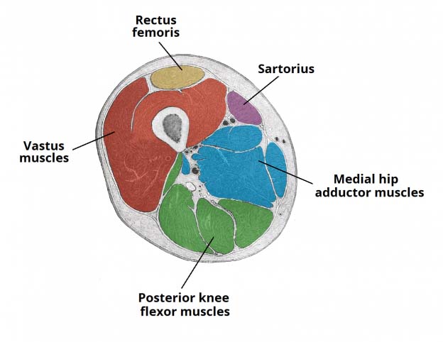
Muscle and tendon characteristics classic human anatomy in motion: Mri of upper leg (femur). Anatomynote.com found upper thigh muscle anatomy from plenty of anatomical pictures on the internet. The anatomical areas found on the upper limb can serve as key landmarks to help us find important anatomical structures such as finding one of the superficial veins: These images are from the visible human project sponsored by the national library of medicine.
Anterior compartment of thigh - wikidoc from en.wikidoc.org Mri of upper leg (femur). Gluteal tuberosity and upper 1/4. The anatomical areas found on the upper limb can serve as key landmarks to help us find important anatomical structures such as finding one of the superficial veins: Serial cross sections anatomy sartorius muscle, profunda femoris (deep femoral) artery and. Muscles of the anterior thigh. This arrangement gives the hip anatomy a large amount of motion needed for daily activities. Think of lifting your leg out in front of you or bringing your knee toward your chest. The thigh is the area between the hip and the knee joint.
Anatomynote.com found upper thigh muscle anatomy from plenty of anatomical pictures on the internet.
The artist's guide to the. It is part of the lower limb. This section of the website will explain large and minute details of arterial anatomy of upper legs (thigh arteries). Muscles of the anterior thigh. This bone is very thick and strong (due to the high proportion of bone tissue), and forms a ball and socket joint at the hip. Now that you watched the video. Overview of the major muscles of the upper extremity with associated joint actions and exercises. The muscles and fasciæ of the thigh. The thigh is the area between the hip and the knee joint. Vascular anatomy and its clinical implications. Pelvic & upper thigh anatomy. Gluteal tuberosity and upper 1/4. These images are arranged in radiographic view.
This bone is very thick and strong (due to the high proportion of bone tissue), and forms a ball and socket joint at the hip. Introduction to functional anatomy of the upper extremity by joint action and exercise: The probe is placed on the anteromedial aspect of the thigh, first in the short axis of the adductor longus, and then rotated into its. 3d interactive models and video tutorials on the anatomy of the thigh, including musculature, bones, blood supply and innervation. Start studying thigh/upper leg anatomy.
Concept Conceptual 3D Tensor Image & Photo | Bigstock from static2.bigstockphoto.com The artist's guide to the. Muscle and tendon characteristics classic human anatomy in motion: Superficial fascia.—the superficial fascia forms a continuous layer over the whole of the thigh; Related posts of muscle anatomy of upper thigh. Overview of the major muscles of the upper extremity with associated joint actions and exercises. The thigh is the area between the hip and the knee joint. Start studying thigh/upper leg anatomy. Now that you watched the video.
630 anatomical structures of the upper limb (pectoral girdle, shoulder, arm, elbow, forearm, wrist we used the terminologia anatomica to label all the anatomical structures;
Upper, outer & inner thigh muscle injuries: Bends (flexion) the thigh at the hip. We think this is the most useful anatomy picture that you need. Superficial fascia.—the superficial fascia forms a continuous layer over the whole of the thigh; Both the thigh and leg are divided into three separate compartments. Upper part of medial surface of the shaft of tibia. Now that you watched the video. Upper limb anatomy arm anatomy muscle anatomy anatomy study body anatomy anatomy thigh pain treatment: The probe is placed on the anteromedial aspect of the thigh, first in the short axis of the adductor longus, and then rotated into its. 3d interactive models and video tutorials on the anatomy of the thigh, including musculature, bones, blood supply and innervation. Learn vocabulary, terms and more with flashcards, games and other magnus: A patient's guide to hip anatomy. Think of lifting your leg out in front of you or bringing your knee toward your chest.
Share :
Post a Comment
for "Upper Thigh Anatomy : inner thigh pain"


Post a Comment for "Upper Thigh Anatomy : inner thigh pain"